Images above acquired on TrueBeam, courtesy of UH Seidman Cancer Center, Cleveland, Ohio
Varian’s HyperSight imaging solution, available as an optional feature on the Halcyon, Ethos, TrueBeam, and Edge radiotherapy systems, allows clinicians to acquire high-quality cone-beam CT (CBCT) images comparable to that of a CT simulation image. The larger images, high soft tissue contrast, and faster image acquisition are designed to improve the ability to target tumor volumes more precisely while sparing healthy tissue.
Below, we’re highlighting key benefits offered by the HyperSight imaging solution’s larger field of view (FOV).
Thanks to the larger kV imaging panel and software innovations that are exclusive with HyperSight, clinicians now have the ability to see more anatomy, in both the axial and transverse planes, that may have previously been cut off or out of view. This may be particularly beneficial when working with larger anatomy, off-center targets, and disease sites where there is extensive nodal involvement. The larger FOV can reduce the need to acquire multiple CBCT images and it may give clinicians the information they need to proceed with off-line adaptation.
The ability to see the entire target is essential for having confidence in the patient's alignment both for imaging and planning, helping enable clinicians to reduce target margins. For patients, this may translate into reduced time on the treatment couch.
Here are examples where the larger FOV translates into clinical impact.
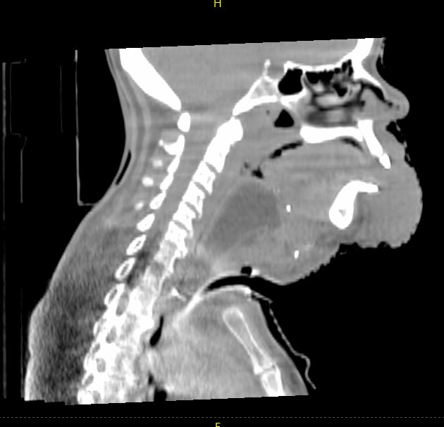
Head and Neck
In this head and neck case with extensive disease involvement, the larger FOV delivered by HyperSight on a Varian TrueBeam system provides greater visibility of the structures and organs surrounding the target to enable a more accurate positional alignment. In this case, the clinician is able to visualize all pertinent anatomy without the need to acquire additional images. This potentially means less time on the treatment couch, less time in the immobilization device, and less imaging dose to the patient.
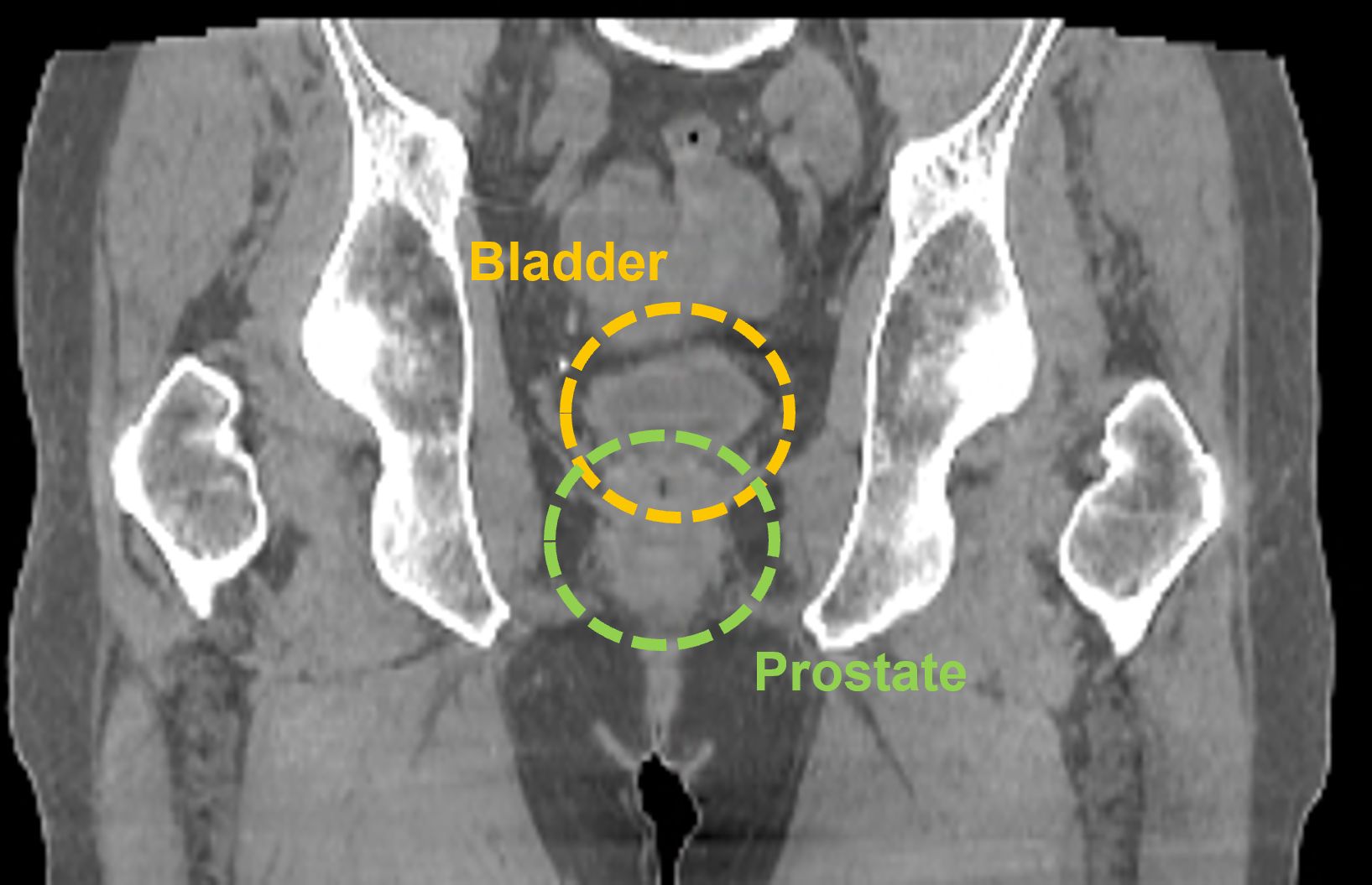
Prostate
In this prostate case, the larger FOV provided by the HyperSight image, taken using a Varian TrueBeam system, enables a clinician to see the entire treatment area including the lymphatic region, which may also require treatment. It also enables the clinician to visualize the relevant anatomy without acquiring multiple CBCT images.
Breast
This HyperSight cone-beam CT for planning (CBCTp*) image, taken using a Varian Halcyon system,** encompasses the entire breast and body, giving clinicians the information they need to generate the initial treatment plan. During treatment, the larger FOV also enables clinicians to see and assess changes in adjacent anatomical structures and landmarks. If the change is deemed significant enough that offline adaptive is needed, they may be able to replan using the latest HyperSight CBCT image.

Learn more about HyperSight:
- TrueBeam With the HyperSight Imaging Solution: The Next Generation of In-Room Imaging
- Foundations of the HyperSight Imaging Solution
- TrueBeam with HyperSight Imaging Solution - The Ohio State Experience
- The Impact to Patients and Clinicians from Implementing the HyperSight Imaging Solution at a Community-based RT Center
*CBCTp is an image-only workflow only available on the Halcyon system.
**HyperSight on Halcyon has different specifications than HyperSight on TrueBeam, as the system configuration for Halcyon allows for a larger kV imaging panel.
QR700020026